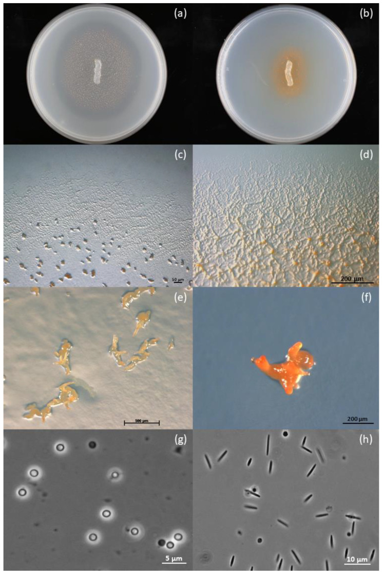
Figure 1.
Growth morphologies of strain ZKHCc1 1396T. (a) Colony on VY/2 agar showing dense orange fruiting bodies around the agar inoculum and lysis of Baker’s yeast cells, as indicated by a halo around it. (b) Colony on CY agar showing darker orange color produced by the swarming cells. (c) Thin and transparent swarm on VY/2 agar with characteristic ripples and flares along the colony edges and fruiting bodies. (d) Swarm on CY agar with pronounced veins and some cell mounds. (e) Coral- or horn-shaped fruiting bodies produced on VY/2 agar. (f) Fruiting body produced on water agar baited with E. coli K-12 bait. (g) Optically refractile and rounded myxospores from a fruiting body produced on water agar. (h) Flexuous and\or slightly tapering vegetative rod cells obtained from CY–H broth. Petri dish diameter is 15 mm (a,b). Stereophotomicrograph (c–f). Phase-contrast photomicrograph (g,h).

