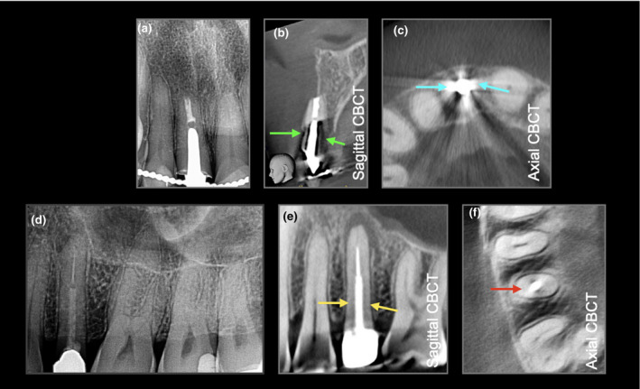
FIGURE 2.

(a) Periapical radiograph and (b, c) sagittal and axial CBCT views assessing a root filled tooth restored with a cast gold post. Note the beam hardening (green arrow) and scatter (cyan arrow). (d) Periapical radiograph of a root filled premolar tooth restored with a fibre post‐retained crown. (e, f) Sagittal and axial CBCT views reveal a periapical radiolucency associated with the tooth; note there is minimal beam hardening (yellow arrow) and scatter (red arrow) with the fibre post
