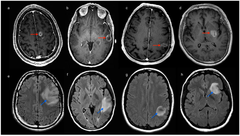
Figure 2.
Images above: post-contrast 3D T1-weighted MR scans of four patients with high-grade glioma (image a–d, red arrows). Tumor location was labeled to account for involved lobes as follows: frontal (a), temporal (b), parietal (c) and insular involvement (d). Images below: FLAIR-weighted MR scans of four patients with low-grade glioma (image e–h, light blue arrows). Tumor location was labeled to account for involved lobes as follows: frontal (e), temporal (f), parietal (g) and insular involvement (h).

