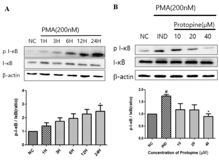
Figure 1.
Protopine regulated I-κBα translocation in PMA-stimulated HepG2 cells. (A) Western blot analysis of time-dependent I-κBα phosphorylation after treatment with PMA 200 nM in HepG2 cells. (B) Cells were pretreated with protopine 40 μM for 1 h and then treated with PMA 200 nM for 24 h. The level of I-κBα protein phosphorylation was determined by Western blot. The results are presented as the means ± SD. # p < 0.05 indicates significantly different from the normal control. * p < 0.05 indicate a significant difference from the PMA group. NC, normal control.

