Figure 3.
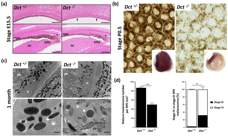
Defect in melanogenesis in the developing RPE of Dct−/− mice. (a) Eosin histology of the E15.5 eye showing the RPE in equatorial (upper panels) and anterior (lower panels) fields; scale bar: 50 μm. Arrows point to unpigmented cells in the RPE of a Dct−/− embryo, whereas at the same stage, all RPE cells are pigmented in the control embryo; Asterisks indicate tip RPE cells with pigment granules located apically in both Dct−/− and control eyes; cmz, ciliary margin zone; nr, neural retina; (b) Bright-field microscopy of P0.5 RPE flat mounts showing a reduced number of pigment granules in Dct−/− samples, scale bar: 10 μm. Inserts show whole eyeballs. At this stage, only the iris and RPE are pigmented in the wild-type, not the outer layer (choroid), so that hypopigmentation of the RPE can be readily detected on enucleated Dct−/− eyes; (c) Transmission electron microscopy of the RPE at 1 month. Low magnification (upper panels, scale bar: 2 μm) shows no difference in the subcellular localization and the shape (round vs. elliptic) distribution of the melanosomes, but these are fewer and less electron-dense in Dct−/− RPE; c, choroid; n, RPE cell nucleus; ps, photoreceptor segment. High magnification (lower panels, scale bar: 500 nm) shows that most of the melanosomes in Dct−/− RPE are at stage III (black arrowheads) or stage IV but with irregular contours (white arrowhead) compared to the controls that are mostly stage IV and exhibit a smooth shape; (d) Quantification of mature melanosomes (left histogram) and stage IV vs. stage III melanosomes (right histogram) from representative sections of one eye per genotype. Total number of analyzed melanosomes: n > 2000 for Dct+/−, n > 1000 for Dct−/−. Values are presented as means ± SEM. Means were compared using t-test, ** p < 0.01; *** p < 0.001.

