Figure 2.
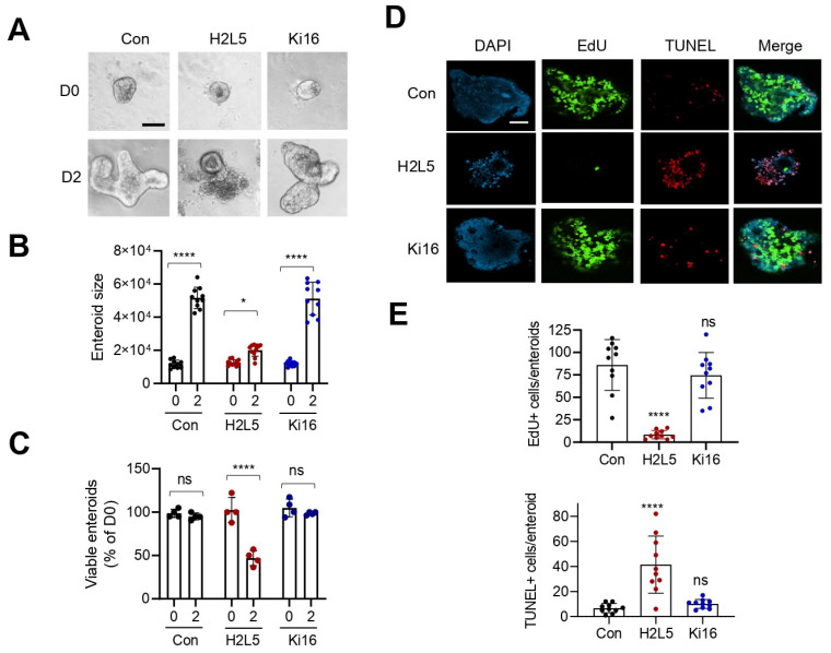
Inhibition of LPA2 in Lpar5f/f;Villi-Cre enteroids results in reduced proliferation and increased apoptosis of IECs. (A) Lpar5f/f;Villi-Cre enteroids were treated with 10 µM LPA2 inhibitor H2L5186303 (H2L5) or LPA1/LPA3 inhibitor Ki16425 (Ki16) on day 0 (D0). Representative images taken on D0 and D2 are shown. n = Bar = 50 µm. (B) The growth of enteroids was quantified by determining the surface area of enteroids using ImageJ. All data are presented as mean ± SD. * p < 0.05, **** p < 0.0001 by two-way ANOVA with Tukey’s multiple comparison test. n = 10. (C) Number of viable enteroids counted on D0 and D2 and expressed as percentage on D0. n = 4 wells per condition. **** p < 0.0001, ns = not significant. (D) Lpar5f/f;Villin-Cre enteroids were treated with H2L5 or Ki. Representative IF images of DAPI (blue), EdU (green), and TUNEL (red) taken on D2 are shown. Bar = 50 µm. (E) Numbers of Edu+ (upper panel) and TUNEL+ (lower panel) cells per enteroid were quantified. n = 10. **** p < 0.0001 and ns = not significant compared with control (Con) by one-way ANOVA with Tukey’s multiple comparison test.

