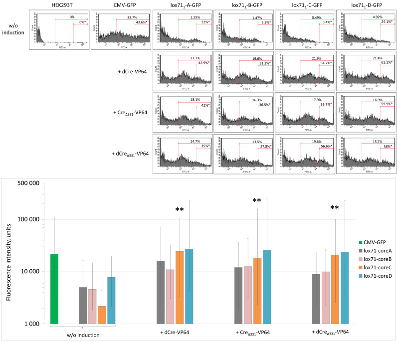
Figure 3.
Flow cytometry analysis of GFP expression in HEK293T cells transfected with various combinations of Cre-VP4/lox71 system components. *, The percentage of bright HEK293T cells among all GFP-positive cells; **, p < 0.05 compared to GFP fluorescence intensity of cells transfected with pVax1-lox711-coreC-GFP plasmid without induction, n ≥ 30,000. In the diagram, data are presented as the median (25%; 75%).

