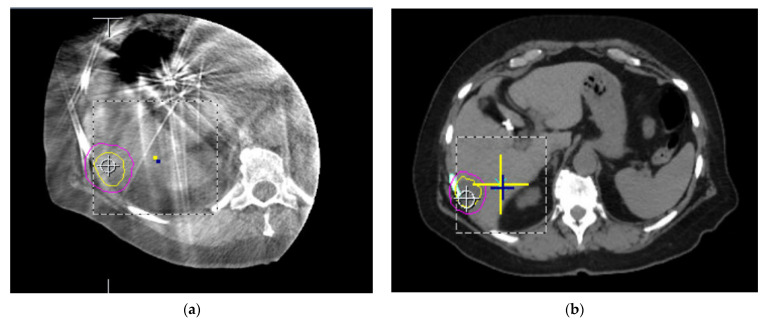
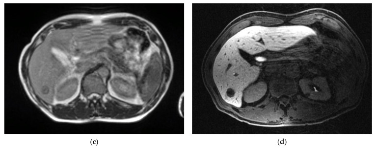
Figure 3.
Comparison of different imaging modalities for the soft tissues of the abdomen. (a) Axial CBCT imaging of the liver taken on a conventional linear accelerator. (b) Axial CT slice of the abdomen used for planning with each organ of a similar electron density. (c) Axial slice of the abdomen acquired with 0.35 T MRI. (d) Axial slice of the abdomen using 1.5 T MRI. Lesion identification is much easier on both 0.35 and 1.5 T MRI images compared to CT and CBCT scans.


