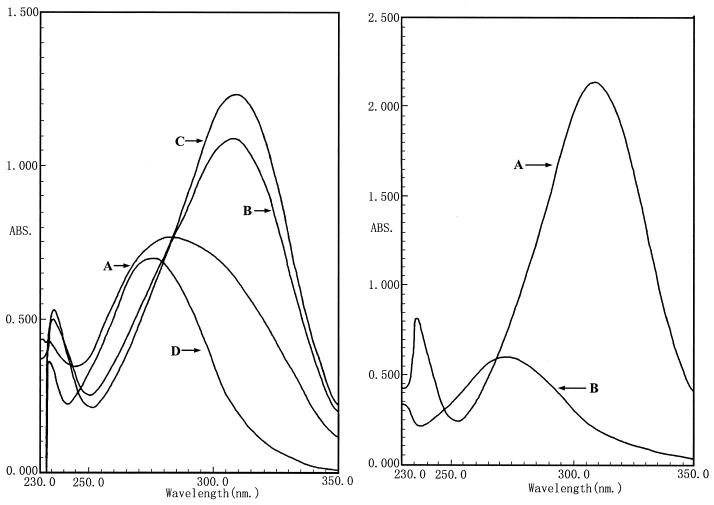
FIG. 1.
(Left) Determination of degradation of methyl parathion by strain M6 at different times by UV scanning. Curves: A, methyl parathion treated with M6 for 5 min; B, treated for 10 min; C, treated for 15 min; D, methyl parathion control. (Right) Localization of the methyl parathion hydrolase enzyme in the cell. Curves: A, methyl parathion treated with the M6 cells; B, methyl parathion treated with supernatant of the culture media. No hydrolase activity was measured in the supernatant.

