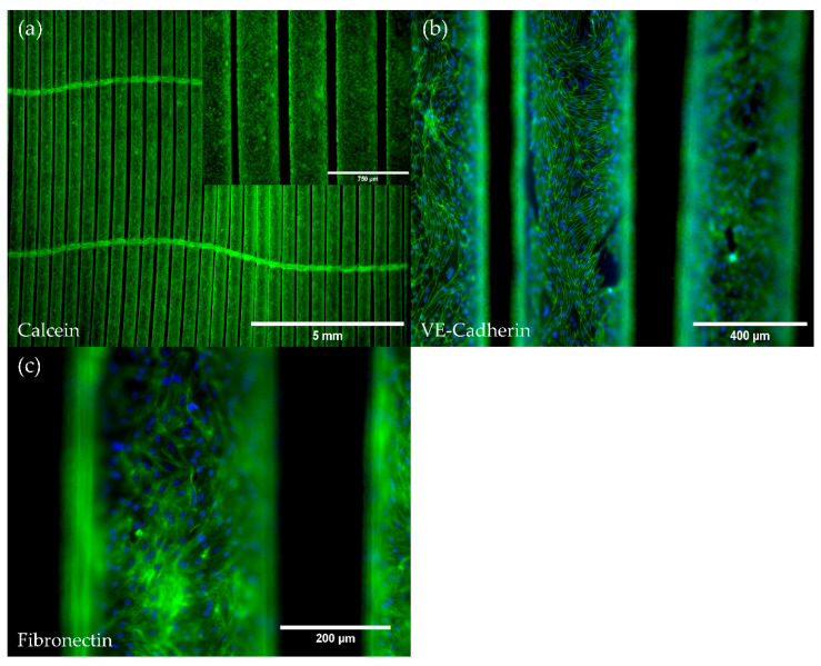
Figure 2.
Viability and immunofluorescence staining of pCECs on HFMs. (a) Calcein staining of viable pCECs (green) forming a confluent monolayer on the HFM; (a) Insert depicts higher magnification; (b,c) Immunofluorescence staining for the detection of (b) VE-Cadherin (green) and (c) the de novo synthesized extracellular matrix protein fibronectin (green); (b,c) Corresponding nuclei were counterstained with Hoechst 33342 (blue).

