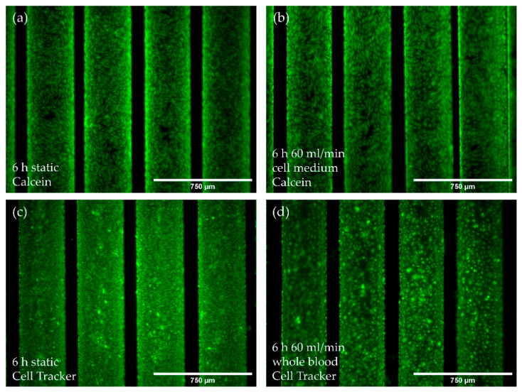
Figure 5.
Analysis of EC monolayer integrity after culture medium or blood flow exposure for six hours. (a,b) Calcein staining (green) of pCECs on HFMs that (a) were kept statically or were exposed to (b) flow conditions using culture medium (60 mL/min); (c,d) Cell Tracker staining (green) of pCECs on statically cultivated HFMs (c) or on 60 mL/min blood flow exposed HFMs (d).

