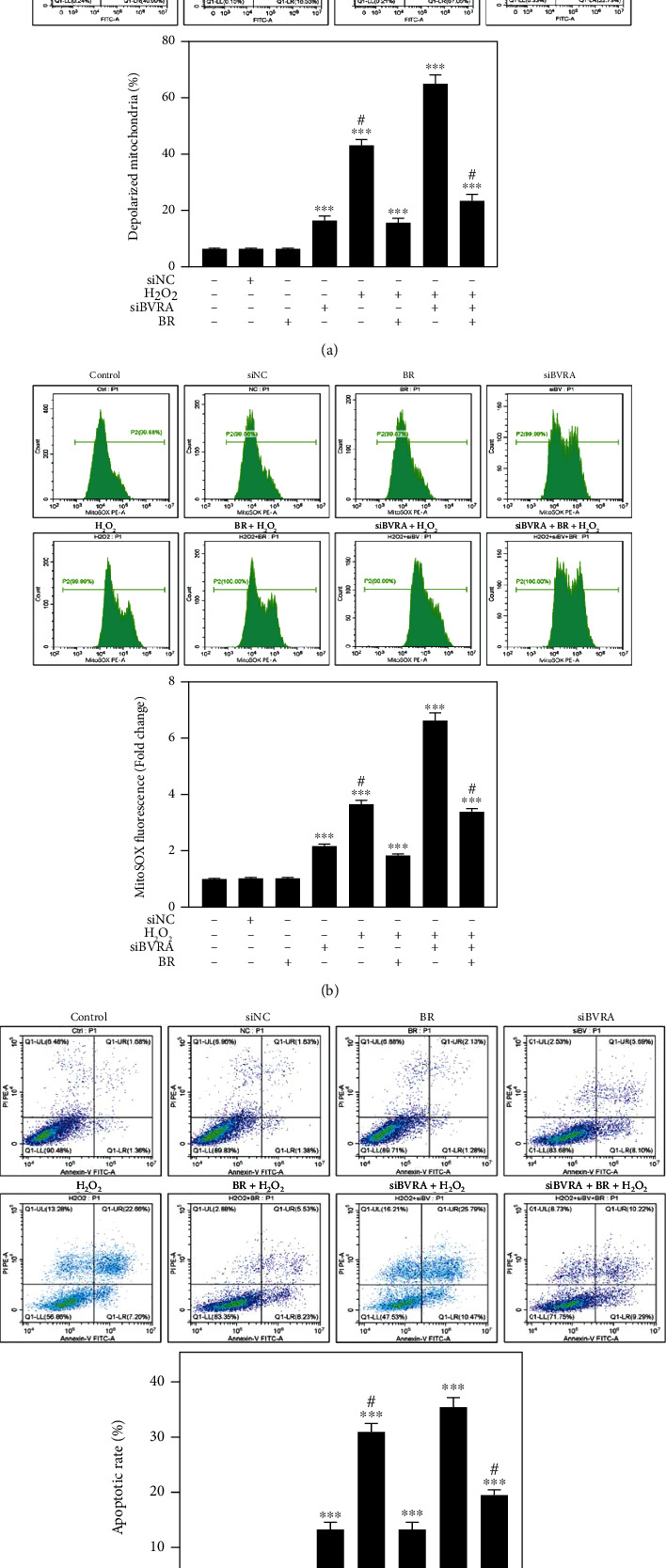
Figure 7.

Mitochondrial dysfunction induced by BVRA depletion under oxidative stress was rescued by BR. LECs were transfected with BVRA siRNA. Then, cells were pretreated with 20 μM BR for 2 h before exposure to 200 μM H2O2 for 24 h. (a) The representative diagram of mitochondrial membrane potential determined by JC-1 staining. (b) Mitochondrial ROS levels were detected by MitoSOX probe. (c) The apoptotic rates of LECs were assessed by Annexin V-FITC assay. Data are shown as mean ± SEM. One-way ANOVA, ∗∗P < 0.01 and ∗∗∗P < 0.001, compared with the control group. #P < 0.05 compared with the BVRA siRNA-transfected cells exposed to H2O2.
