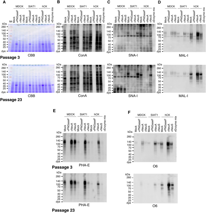
Figure 4.
Lectin and Western blot analyses of total glycoproteins in MDCK, SIAT1, and hCK cells. Total cell lysates of MDCK, SIAT1, and hCK cell lines were treated with PNGase F (PNGaseF), Neuraminidase A (NeuA), or Neuraminidase S (NeuS), and separated by SDS-PAGE. Total extracts of cells at passage 3 (top) and passage 23 (bottom) (A–F), plus PNGaseF/neuraminidases mixture (Enzyme mix) control were loaded on the gels. (A) Gel stained with Coomassie Brilliant Blue solution. (B) Transferred total glycoproteins analyzed by lectin blot using ConA; (C) SNA; (D) MAL-I; and (E) PHA-E. (F) Total glycoproteins from cells at passage 3, and 23 were Western blotted with the antibody O6.

