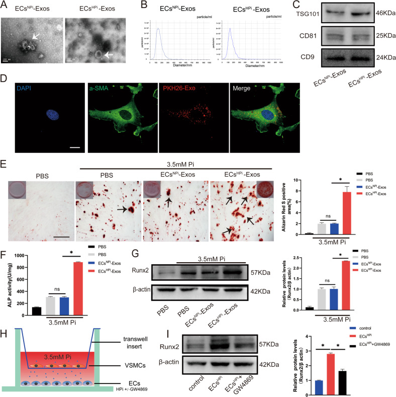
Fig. 2. Exosomes secreted from high phosphorus-stimulated ECs exacerbated VSMCs calcification.
A Transmission electron micrographs of exosomes derived from ECsNPi-Exos or ECsHPi-Exos phosphate conditions (Bar = 100 nm). B The nanoparticle concentration and size distribution of the ECs-Exos. C CD9, CD81, and TSG101 immunoblots of exosomes. D VSMCs were incubated with PKH26 fluorescently labeled exosomes for 12 h. Confocal microscopy analysis was used to identify the uptake of EVs by VSMCs (PKH26 in red, DAPI in blue, and α-SMA in green) (Scale bar = 5 μm). E VSMCs were incubated with ECs NPi-Exos and ECsHPi-Exos for 48 h, respectively. Then, Alizarin Red S staining was performed and calcium content was measured in VSMCs. The black arrows indicate mineralized nodules in VSMCs (Scale bar = 200 μm). F ALP activity was measured by an ALP kit in VSMCs incubated with ECsNPi-Exos and ECsHPi-Exos. G The expression of Runx2 was determined by Western blot in VSMCs incubated with ECsNPi-Exos and ECsHPi-Exos. H, I ECs in the lower chamber pre-treated with or without GW4869 were co-cultured with VSMCs in six-well Transwell units, and the upper chamber VSMCs were harvested to determine the expression of Runx2 by using western blot. Three independent experiments were performed, and representative data were shown. Data are shown as mean ± SD. ns: no significance, *p < 0.05.

