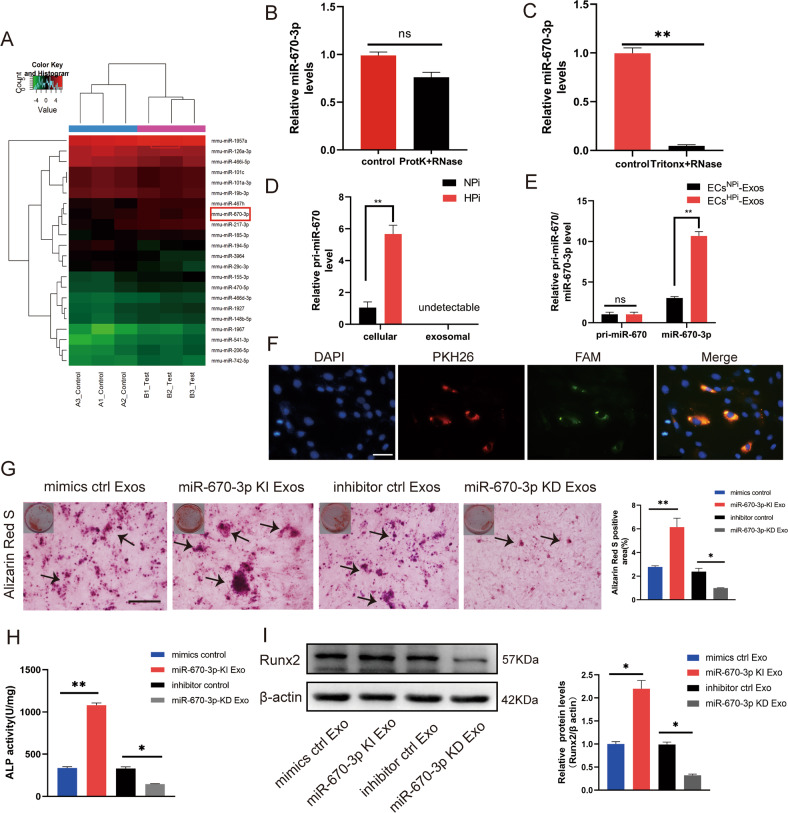
Fig. 3. miR-670-3p was enriched in ECsHPi-Exos and regulated VSMCs calcification.
A The differentially expressed miRNAs (a cut-off of absolute fold change ≥1.5 and p < 0.05) between ECsNPi-Exos and ECsHPi-Exos according to microarray analysis. B qRT-PCR quantitative results of miRNA after exosomes were treated with proteinase K and RNase. C qRT-PCR quantitative results of miRNA after exosomes were treated with Tritonx-100 and RNase. D Expression levels of pri- miR-670-3p in ECs and EC-Exos. E Expression levels of pri-miR-670-3p and mature miR-670-3p in VSMCs. F Confocal microscopy analysis was used to verify whether exosomal miR-670-3p could be uptaken by VSMCs. VSMCs were cultured with PKH26-labeled exosomes derived from ECs transfected FAM-miR-670-3p-mimics. The FAM-miR-670-3p signals were detected in the cytoplasm of VSMCs (Green), and FAM-miR- 670-3p signals were co-localized with PKH26 in VSMCs (PKH26 in red and DAPI in blue), (scale bar = 50 μm). G–I VSMCs were incubated with miR-670-3p knocked-in exosomes or miR-670-3p knocked-down exosomes during a high phosphorus induction period. Then, the severity of VSMC calcification was evaluated by Alizarin Red S staining (G), ALP activity (H), and Runx2 protein expression (I). The black arrows (G) indicate mineralized nodules in VSMCs (scale bar = 100 μm). Three independent experiments were performed, and representative data were shown. Data were shown as mean ± SD. ns: no significance, *p < 0.05, **p < 0.01. ns: no significance.

