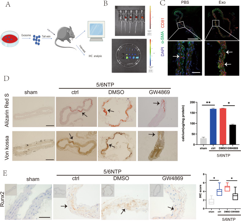
Fig. 5. ECs-Exos could be internalized by the artery and mediated arterial calcification.
A Schematic representation of procedure to trace ECs-Exos in mice. The ECs-Exos were labeled with the membrane-philic dye DIR and injected into mice through the tail vein. PBS was used as a negative control, and DIR dye was used as a positive control (n = 5 per group). B After 24 h, fluorescence signals were detected in the living mice (upper panel) and their organs after execution (lower panel). C The thoracic aorta was obtained to analyze the uptake of endothelial exosomes in the slices. Blue fluorescence (DAPI)-labeled cell nuclei, green fluorescence (Alexa 488)-labeled α -SMA, and red fluorescence (Alexa 555)-labeled CD81 indicating exosomes(The upper scale bar = 200 μm, the lower scale bar = 20 μm). The white arrow indicates CD81 positive exosomes. D, E Evaluation of the effect of pre-treatment of the exosome blocker GW4869 on arterial calcification induced by 5/6 nephrectomy. D Alizarin Red S (upper channel) and Von Kossa (lower channel) staining analysis of paraffin-embedded mouse vascular tissues. The arrows indicate mineralized nodules (scale bar = 200 μm). E Immunochemistry analysis of osteogenic differentiation marker Runx2 expression. The arrows indicate the positive expression of Runx2 in the mouse artery (scale bar = 50 μm). Data are represented as the mean ± SD, with five replicates for each group. Two-way ANOVA analyzed significance with Turkey’s HSD post hoc analysis. *p < 0.05, **p < 0.01.

