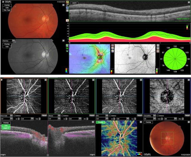
Fig. 1.
OCT Topcon ImageNet 6 (DRI OCT Triton, Topcon Corporation) was used to obtain OCT swept-
source angiography scans from an area of 4.5 × 4.5 mm2 centered on the optic nerve. Four layers were considered: superficial capillary plexus (SCP), deep capillary plexus (DCP), outer retina (OR) and choriocapillaris (CC). ImageJ software was used to calculate the peripapillary vascular index. Pictures were converted from black and white into binary images; afterward, colors channel was adjusted through “color threshold” in order to visualize vascular structures. Peripapillary region was obtained by drawing two ellipses around it (270X230 nm and 60X60 nm), visualizing the Vascular Index of the interested area

