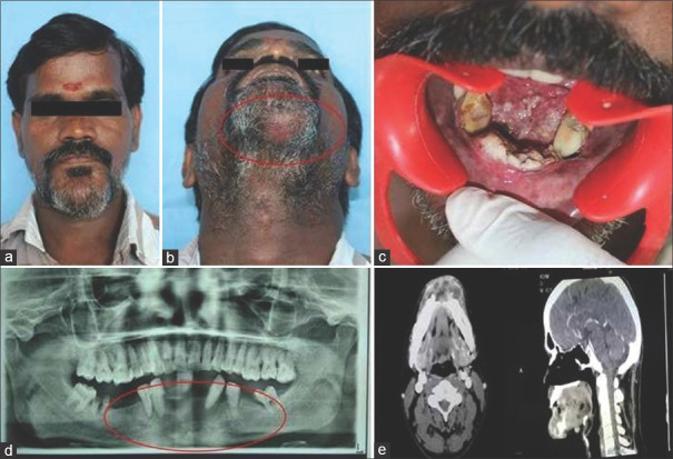
Figure 1.
Preoperative pictures (from right top to down). (a and b) Extraoral clinical picture showing skin induration, (c) Intraoral picture of ulceroinfiltrative lesion involving floor of the mouth and lower alveolus, (d) Orthopantomogram, (e) CECT scan of the head and neck with axial and sagittal view

