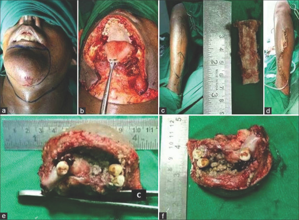
Figure 2.
Intra operative pictures (from right top to down). (a) Incision marking and (b) post excision defect, (c) harvestment of nonvascularized bone graft of fibula of 6.5 cm from left leg (d) closure of donor site with drain, (e and f) Specimen picture of full thickness wide excision of skin of chin with anterior segmental mandibulectomy and floor of the mouth with adequate margin

