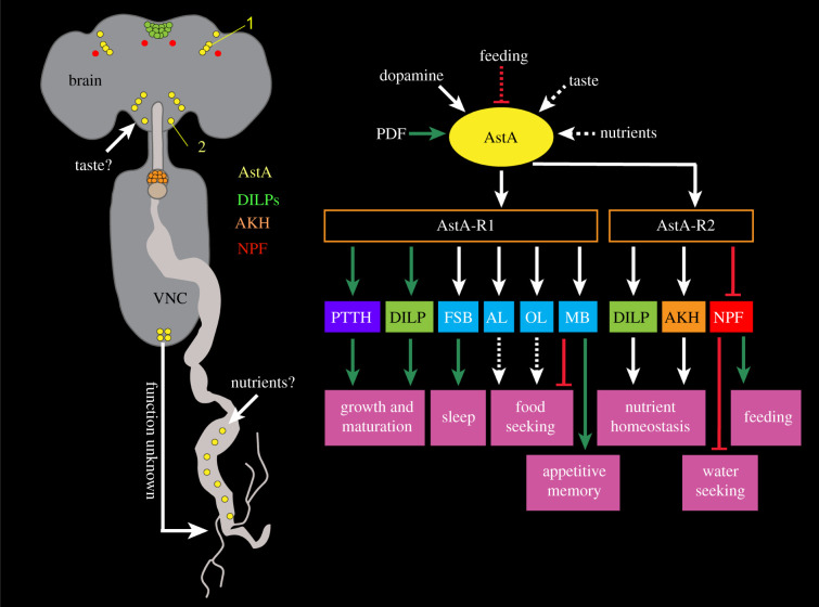
Figure 6.
Context-specific signalling with Allatostatin-A (AstA). AstA neuropeptide from the brain and gut regulates diverse feeding associated behaviours and physiology. (a) A schematic showing the locations of select AstA-expressing cells in the nervous system and midgut, as well as some of its downstream neuronal targets. The morphology and location of AstA cells suggest that the suboesophageal zone AstA neurons may receive taste inputs from the proboscis, and AstA-expressing enteroendocrine cells in the gut may be nutrient-sensitive. A PLP neuron is indicated by 1 and a Janu-AstA by a 2; these are discussed in the text. (b) Inputs and behavioural outputs of AstA cells. AstA-expressing neurons receive inputs from the pigment-dispersing factor (PDF) expressing clock neurons [91] and dopaminergic inputs via the Dop1R1 receptor [9]. In addition, feeding inhibits AstA neurons and they may also receive gustatory inputs via the proboscis and nutrient information via the gut. AstA, in turn, mediates its effects via its two receptors, AstA-R1 and AstA-R2 expressed in various peptidergic cells/neurons. These include cells expressing prothoracicotropic hormone (PTTH), insulin-like peptides (DILPs), adipokinetic hormone (AKH) and neuropeptide F (NPF). AstA-R1 is also expressed in neurons in the fan-shaped body (FSB) and mushroom body (MB). Moreover, axonal projections of AstA neurons to the antennal lobe (AL) and optic lobe (OL), coupled with single-cell transcriptome data from the Fly Cell Atlas suggest expression of AstA-R1 in the AL and OL. AstA modulation of peptidergic neurons and other neuronal targets influences various feeding-related behaviours including feeding, appetitive memory and water seeking. Green arrows represent stimulation, red bars represent inhibition, white arrows represent unclear valence and dashed arrows represent postulated actions. (b) is based on [9,20,91,92,180–183].

