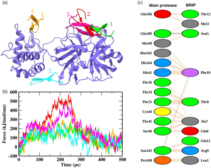
Figure 3.
Detection of the protein-peptide binding site. (a) BRIP docked on the top five predicted binding sites on Mpro. (b) The pull force profiles of BRIP attached to the binding pockets of Mpro. The pull force trajectories are colored according to the binding poses shown in panel a. (c) The Mpro-BRIP interactions at the most favorable binding site. The solid blue color lines represent hydrogen bonds, while the striped lines denote non-bonded interactions. The residue color coding scheme: positive (blue), negative (red), neutral (green), aliphatic (gray), aromatic (purple), proline and glycine (orange).

