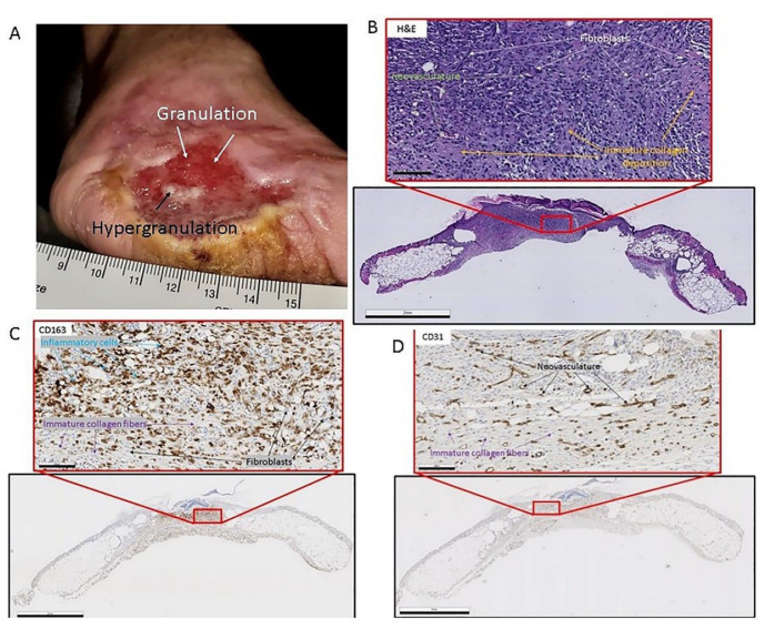
Figure 2.
(A) Clinical image of chronic diabetic injury with granulation tissue presence. The wound is on the medial aspect of the foot. Notice the white arrow pointing to red tissue indicating cellular infiltrate and neovascularization. The black arrow is pointing to the area in the middle of the wound which is indicative of hyper-granulation. (Patient consent provided). (B) Heamatoxalin and Eosin-stained murine skin the black frame is a gross image (scale bar = 2 mm) of the skin sample, and the red framed image (scale bar = 100 µm) is a magnified image of the area in the small red box. The white arrows point to fibroblasts in the granulation tissue. The orange arrows point to immature collagen deposition. The green arrows point to neovasculature. (C) CD-163 (Inflammatory cell marker) reacted immunohistochemistry-stained murine skin the black frame is a gross image (scale bar = 2 mm) of the skin sample and the red framed image (scale bar = 100 µm) is a magnified image of the area in the small red box. The black arrows point to fibroblasts in the granulation tissue. The purple arrows point to immature collagen deposition. The blue arrows point to inflammatory cells: macrophages, and monocytes. (D) CD-31 (endothelial cell marker) reacted immunohistochemistry-stained murine skin, the black frame is a gross image (scale bar = 2 mm) of the skin sample, and the red framed image (scale bar = 100 µm) is a magnified image of the area in the small red box. The black arrows point to neovasculature in the granulation tissue. The purple arrows point to immature collagen deposition.

