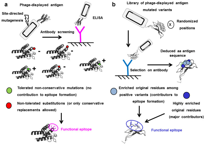
Figure 2.
Epitope identification through mutagenesis scanning of the antigen surface. (a) Phage-displayed antigen is diversified through site-directed mutagenesis targeting a candidate antigenic area previously identified. Single mutated variants are tested by ELISA with the antibody under investigation, and classified as positive or negative. Those positions that cannot be mutated without abolishing antigen recognition (or where only conservative replacements are accepted) are defined as functional contributors to the epitope. (b) Combinatorial mutagenesis within a candidate antigenic region results in construction of a library of multiple mutated antigen variants. Phage panning on an immobilized antibody leads to selection of recognized (positive) variants. Sequencing reveals that several original residues are enriched to different extents among them, and the whole cluster is thus identified as the functional epitope.

