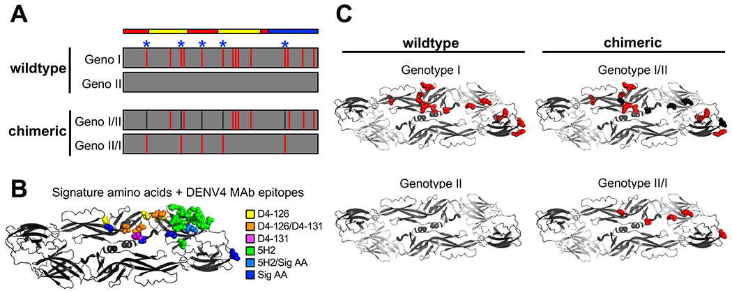
Figure 4. Design of GI and II chimeric viruses for mapping key residues.

A) Schematic of E protein amino acid differences between DENV4 GI virus relative to GII virus. The figure depicts signature amino acids (blue asterisks), and design of chimeric viruses, aligned with envelope protein domains (red = EDI, yellow = EDII, blue = EDIII). B) Signature amino acids and DENV4 MAb epitopes shown on one monomer within the E protein dimer. C) Amino acid differences of GI (red) relative to DEVN4 GII, and signature amino acids transplanted in chimeric viruses (black and red) (PDB = 1OAN).
