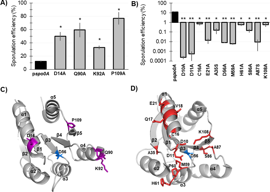
Figure 3. Mutagenesis of conserved Spo0A residues results in both increased and decreased C. difficile sporulation frequency.

A) Ethanol-resistant spore formation of 630Δerm spo0A pspo0A (MC848) expressed on a plasmid compared to the Spo0A site-directed mutants D14A (MC1671), Q90A (MC1712), K92A (MC1185), and P109A (MC1621) with increased sporulation frequency. B) Ethanol-resistant spore formation of 630Δerm spo0A pspo0A (MC848) expressed on a plasmid compared to the Spo0A site-directed mutants D10A (MC1618), D11A (MC1703), C16A (MC1057), E21A (MC1058), A35S (MC1059), D56A (MC849), M59A (MC1184), H61A (MC1036), S86A (MC1846), A87S (MC1061), and K108A (MC1064) with decreased sporulation frequency, displayed on log10 scale. Sporulation assays were performed independently at least four times. Statistical significance was determined using Kruskal-Wallis test and uncorrected Dunn’s test (*, P = > 0.05; **, P = > 0.01). C) 3D structure of Spo0A with residues (highlighted purple) that cause increased sporulation when mutated, orientated around the activation site (D56, highlighted blue). D) 3D structure of Spo0A with residues (highlighted red) that reduce sporulation when mutated, orientated around the active site (D56, highlighted blue). Spo0A PDB code 5WQ0, edited in PyMOL (The PyMOL Molecular Graphics System, Version 2.4.0 Schrödinger, LLC).
