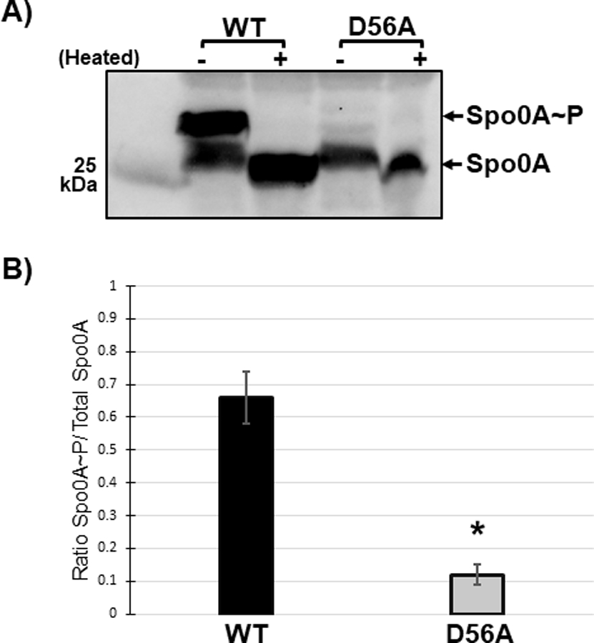
Figure 4. The conserved aspartate residue of C. difficile Spo0A is phosphorylated.

A) Anti-FLAG western blot after phos-tag gel separation of unphosphorylated and phosphorylated Spo0A (Spo0A~P) species in 630∆erm spo0A pspo0A-3XFLAG (MC1003) and 630∆erm spo0A pspo0A D56A-3XFLAG (MC1690) grown on sporulation agar. Phos-tag SDS-PAGE was performed on protein extracts (10 ug) and visualized using an anti-FLAG antibody. The molecular weight marker (25 kDa) is indicated on the left of the panel and experiments were performed 3 independent times. B) Ratio of phosphorylated Spo0A to total Spo0A. Densitometry calculations were performed using ImageJ 1.53a. (*, P = < 0.01) as determined by unpaired two-tailed Student’s t-test.
