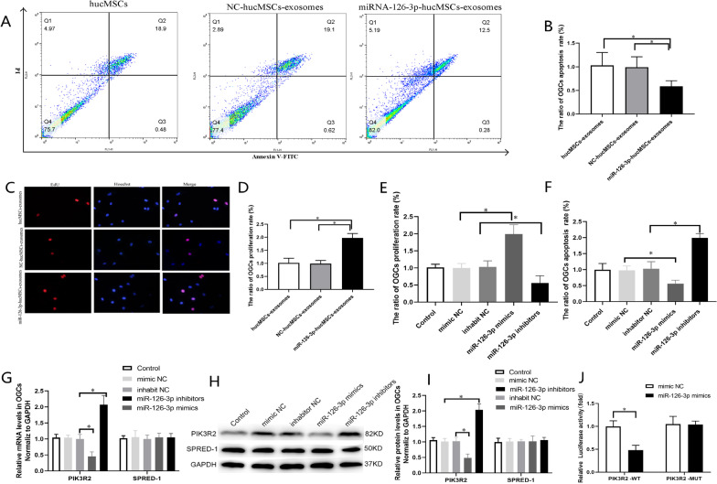
Fig. 3.
miR-126-3p promoted proliferation and inhibited apoptosis of OGCs in vitro and PIK3R2 is a target of miR-126-3p. A. Flow cytometric apoptosis assay was used to analyze the effect of miR-126-3p-hucMSCs-exosomes on OGCs apoptosis. B. Analysis of the ratio of the percentages of apoptotic OGCs in each group. C. EDU assay was used to analyze the effect of miR-126-3p-hucMSCs-exosomes on OGCs proliferation. D. Analysis of ratio of the percentages of proliferative OGCs in each group. E. Comparison of the ratio of apoptosis rate of OGCs as flow cytometric apoptosis assay in each group. F. Comparison of the ratio of proliferation rate of OGCs as EDU assay in each group. G. The mRNA levels of PIK3R2 and SPRED-1 after transfection of miR-126-3p mimics and inhibitor in OGCs. H. Representative western blots of PIK3R2 and SPRED-1 protein expression in OGCs transfected with miR-126-3p mimics and inhibitor. I. The protein levels of PIK3R2 and SPRED-1after transfection of miR-126-3p mimics and inhibitor in OGCs. J. After transfection of miR-126-3p mimic and wild-type or mutant vectors into OGCs, dual-luciferase reporter genes were performed to determine luciferase activity**P < 0.01 versus WT group. *P < 0.05 for all figures

