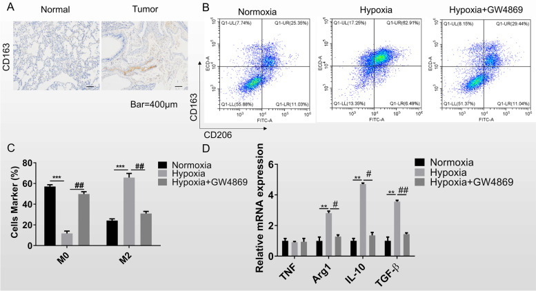
Fig. 1.
Hypoxic lung cancer cells promote macrophage M2 polarization. A IHC was performed to quantify the CD163 protein expression in clinical tissues. Scale bar = 400 μm. B Flow cytometry was used to detect the quantity of M2 macrophage through labeling the protein CD206 and CD163. GW4869 was used as the inhibitor of exosome secretion. C The quantitative analysis of flow cytometry results. D RT-qPCR was for the marker genes examining in mTHP-1 that co-cultured with the normoxia and hypoxia lung cancer cell H1299. Data was shown as mean ± SD. n = 3. *P < 0.05, **P < 0.01, ***P < 0.001

