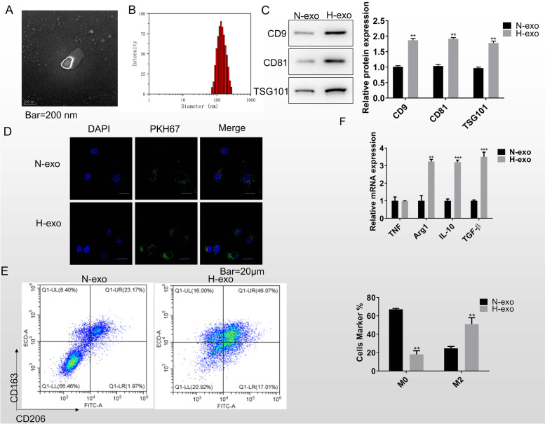
Fig. 2.
Exosomes from hypoxic lung cancer cells promote macrophage M2 polarization. A Exosome morphological characteristics were determined by transmission electron microscope. Scale bar = 200 nm. B Dynamic light scattering was used to detect the exosome size. C The expression of exosome protein biomarkers CD9, CD81, and TS101 was measured by western blot. D The immunofluorescence image shows the internalization of PKH67-labeled exosomes by macrophage. Scale bar = 20 μm. E Flow cytometry was used to detect the quantity of CD163+CD206+ M2 macrophage after treated with normoxia or hypoxia exosome for 48 h. F RT-qPCR was for the marker genes examining in macrophages that co-cultured with the normoxia and hypoxia exosomes, including TNF, Arg1, IL-10, and TGF-β. Data was expressed as mean ± SD. n = 3. *P < 0.05, **P < 0.01, ***P < 0.001

