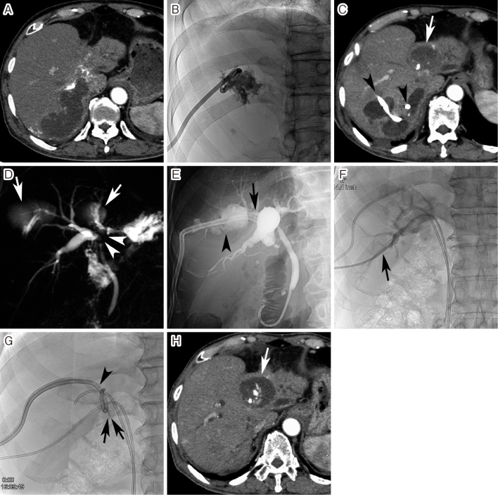
Fig. 5.
Embosclerosis of intrahepatic biloma
A. Arterial-phase computed tomography (CT) performed 1 month after transarterial chemoembolization (TACE) showed a large biloma in the posterior segment of the right hepatic lobe. B. Percutaneous transhepatic biloma drainage was performed. C. Bile discharge from the biloma continued despite multiple ablation sessions using 5% ethanolamine oleate. CT performed 3 months after TACE showed the development of multiple bilomas in the right hepatic lobe. The arrowheads indicate the drainage catheter passing through three bilomas. The arrow also indicates a previously embolized tumor 9 years and 8 months ago. D. Magnetic resonance cholangiopancreatography showed bile duct strictures (arrowheads) at the hepatic hilum due to TACE, as well as the bilomas (arrows). E. A plastic endoprosthesis was endoscopically placed into the anterior segmental bile duct (arrow). However, the endoprosthesis could not be advanced into the posterior segmental bile duct branch. The biloma was also demonstrated (arrowhead). F. A drainage catheter was percutaneously advanced into the posterior segmental bile duct branch (arrow). G. Thereafter, a fibrin glue was injected into the biloma. The arrows indicate two 4-F catheters advanced into the biloma cavity. The arrowhead also indicates the drainage catheter in the biloma. Four sessions of the fibrin glue injection were performed at 3–4-day intervals. Thereafter, bile discharge was stopped and the drainage catheter could be removed. H. Arterial phase CT performed 4 months after embosclerosis of biloma showed the disappearance of all bilomas. The arrow indicates the previously embolized tumor.

