Figure 3:


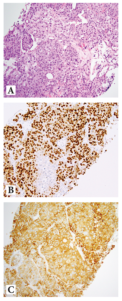
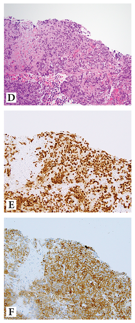
Prostate cancer with diffuse neuroendocrine differentiation
Example 1: (A) H&E at 10x magnification highlighting nested/compartmentalized arrangement of cells with abundant amphophilic cytoplasm; (B) diffuse reactivity with NKX3.1, and (C) diffuse reactivity with synaptophysin
Example 2: (D) H&E at 10x magnification showing sheet-like growth of cells with abundant amphophilic cytoplasm; (E) diffuse reactivity with NKX3.1, and (F) diffuse reactivity with synaptophysin
