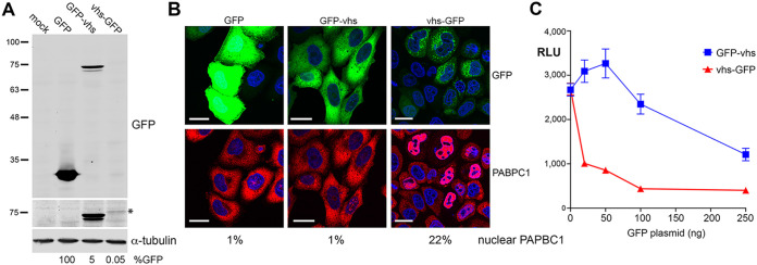
FIG 1.
Characterization of GFP-tagged vhs proteins. (A) HeLa cells were mock-transfected or transfected with plasmids expressing GFP-tagged proteins as shown. After 20 h, total cell lysates were analyzed by Western blotting for GFP and α-tubulin. Relative expression of GFP was quantitated using LICOR ImageStudio and is represented as a percentage of untagged GFP level, normalized to tubulin (B). (A) Transfected cells grown on coverslips were fixed and stained for PABPC1 (red) and nuclei stained with DAPI (blue). Scale bar = 20 μm. The percentage of cells in the monolayer with nuclear PABPC1 is shown (>200 cells counted). (C) HeLa cells grown in 24-well plates were transfected with 50 ng plasmid expressing Gaussia luciferase, together with increasing amounts of a plasmid expressing either GFP-vhs or vhs-GFP. After 16 h, the medium was changed and 6 h later the medium was sampled, and relative luciferase levels were measured by injection of coelenterazine in a Clariostar plate reader. The means ± standard errors of the means (SEM) for one representative experiment are shown (n = 4). RLU, relative light units (arbitrary).

