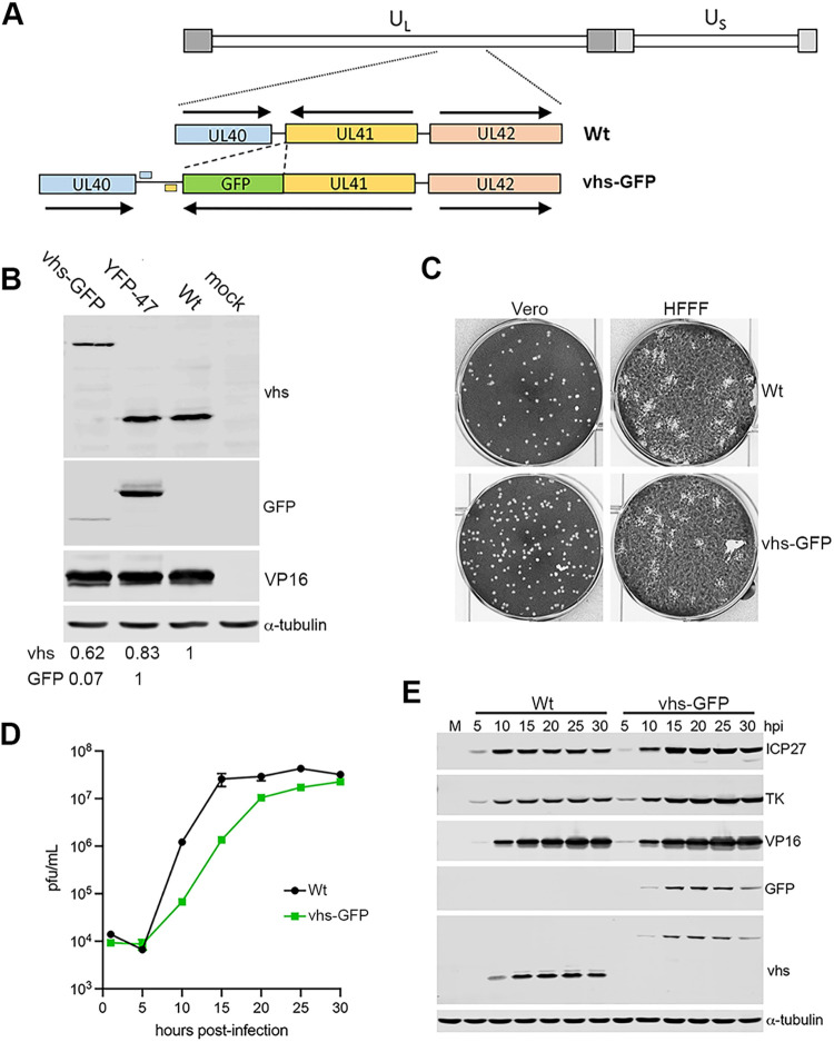
FIG 3.
Construction and characterization of HSV1 expressing vhs-GFP. (A) HSV1 expressing vhs-GFP in place of GFP was constructed by cotransfecting Vero cells with infectious Sc16 genomic DNA and a plasmid containing the UL41GFP fusion gene surrounded by the flanking sequences from UL40 and UL42. Polyadenylation signals for UL40 and UL41 are shown. (B) HFFF cells were mock-infected or infected with Sc16, plaque purified vhs-GFP or YFP-UL47 viruses at an MOI of 2, and total protein harvested at 16 hpi before separation by SDS-PAGE and Western blotting with antibodies for GFP, vhs, VP16, and α-tubulin. Relative expression of vhs and GFP were quantitated using LI-COR ImageStudio and normalized to α-tubulin. (C) Vero and HFFF cells were infected with around 40 PFU of Sc16 or vhs-GFP and incubated for 3 days before fixation and staining with crystal violet. (D) One-step growth curves for WT (Sc16) and HSV1 vhs-GFP viruses were carried out by harvesting the total virus from HFFF cells infected at an MOI of 2 every 5 h up to 30 h. Samples were titrated onto Vero cells. The mean titer of three replicates and associated SEM are plotted. (E) Total extracts of HFFF cells infected as in (D) were analyzed by SDS-PAGE and Western blotting for the indicated virus proteins and α-tubulin as a loading control.

