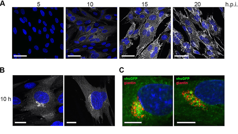
FIG 4.

Localization of vhs-GFP during HSV1 infection. (A) HFFF cells infected with HSV1 vhs-GFP at an MOI of 2 were fixed at the indicated times, stained with DAPI (blue), and imaged for GFP fluorescence (white) using confocal microscopy. Scale bar = 50 μm. (B) Examples of the 10 h sample from (A) imaged at higher magnification. Scale bar = 20 μm. (C) HFFF cells infected as in (A) were fixed and stained for the Golgi marker giantin. Scale bar = 10 μm.
