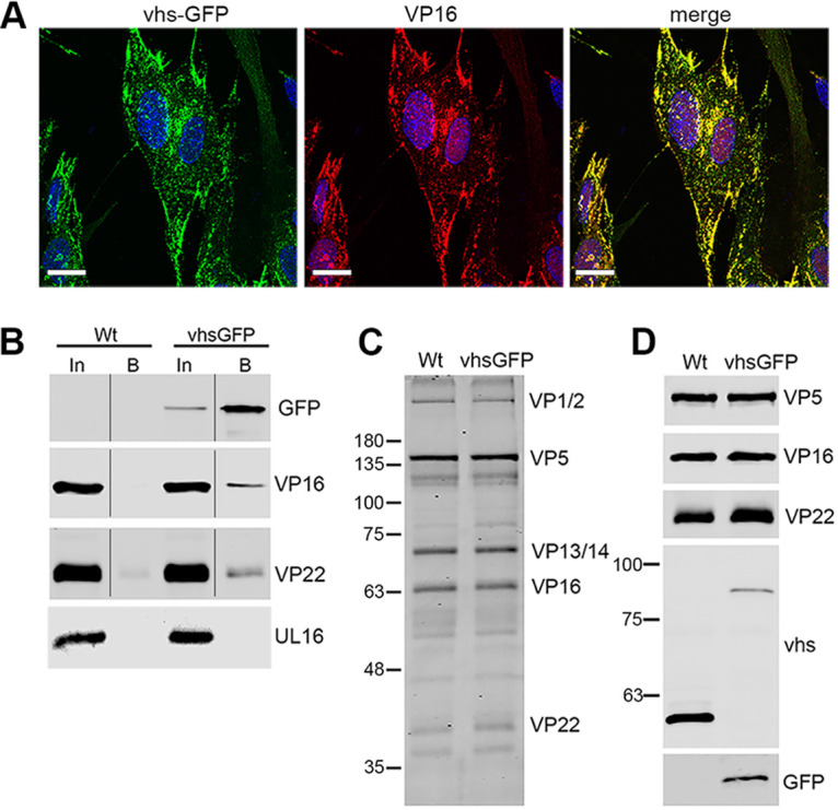
FIG 5.

Interaction of vhs-GFP with VP16 and assembly into virions. (A) HFFF cells infected with HSV1 vhs-GFP at an MOI of 2 were fixed at 16 h, stained for VP16 (red), and imaged for GFP (green). Nuclei were stained with DAPI (blue). Scale bar = 20 μm. (B) GFP-Trap pulldown was carried out on HaCat cells infected with WT (Sc16) or vhs-GFP at an MOI of 5 and harvested at 24 hpi. The pulldowns were analyzed by SDS-PAGE and Western blotting with antibodies to GFP, VP16 and VP22, and UL16 as a control for nonspecific pulldown of virus protein. In = input, B = pulldown (C and D) Extracellular virions were purified on 5% to 15% Ficoll gradients and analyzed by SDS-PAGE followed by Coomassie blue staining (C) or Western blotting with antibodies as indicated (D).
