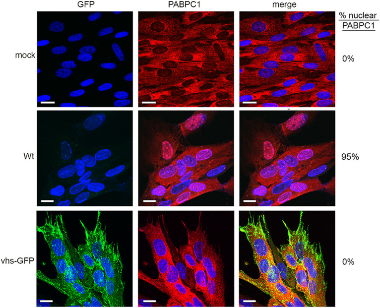
FIG 6.

PABPC1 is not relocalized in HSV1 vhs-GFP infection. HFFF cells infected with WT (Sc16) or HSV1 vhs-GFP viruses at an MOI of 2 were fixed at 16 h, stained with an antibody for PABPC1 (red), and nuclei stained with DAPI (blue). Scale bar = 20 μm. Percentage cells in the monolayer with nuclear PABPC1 are shown on the right-hand side.
