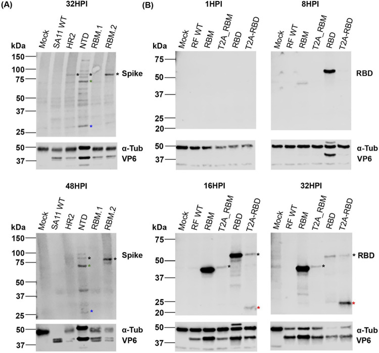
FIG 4.
Expression of VP6 and SARS-CoV-2 spike peptides in infected cells. Cells infected at low MOI were harvested at 1, 8, 16, 24, 32, and 48 hpi. Whole-cell lysates were analyzed by SDS-PAGE and Western blot using polyclonal antibodies against RV VP6, and spike for SA11 mutants (A) or RBD for RF mutants (B). Alpha-tubulin (α-Tub) was used as a loading control. In panel A, black asterisks denote uncleaved VP4 product. Cleaved VP4 products VP5* and VP8* are marked by green and blue asterisks respectively. In panel B, black asterisks show T2A read-through product and red asterisks identify separated products. Representative results from three independent experiments are shown. Position of molecular weight markers are indicated (kDa).

