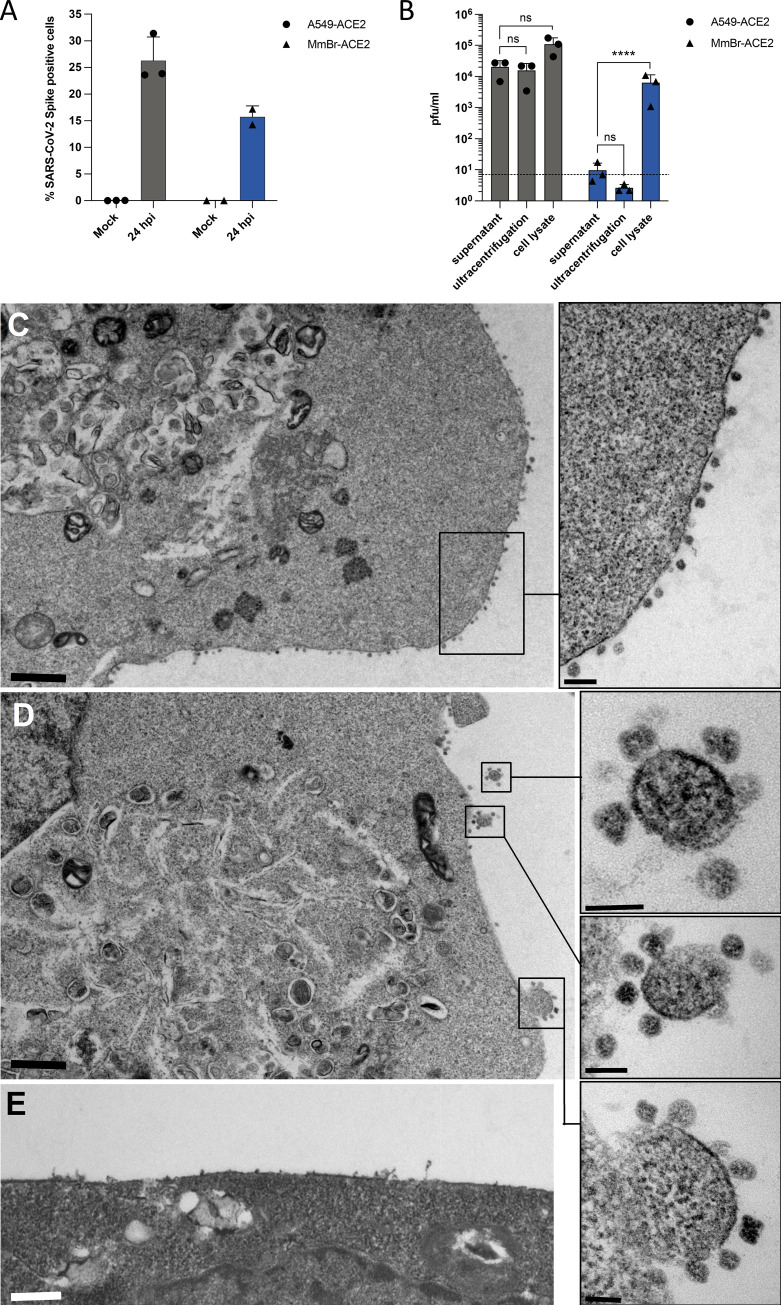
FIG 6.
Infectious particles are retained at the surface of MmBr-ACE2 cells. A549-ACE2 and MmBr-ACE2 cells were left uninfected (Mock) or were infected for 24 h at an MOI of 1 or 0.04, respectively. (A) The percentages of cells that contained SARS-CoV-2 S proteins were determined by flow cytometric analysis. (B) The presence of extracellular infectious viruses in the culture medium of the cells was determined by TCID50 assays performed on Vero E6 cells. Supernatants were either clarified or clarified and purified by ultracentrifugation. Alternatively, cell-associated infectious virions were titrated on Vero E6 cells from whole-cell lysates. Data points represent three independent experiments. Statistical test: Dunnett’s multiple-comparison test on a two-way ANOVA (ns, not significant; *, P < 0.05; **, P < 0.01; ***, P < 0.001; ****, P < 0.0001). (C to E) At 16 h postinfection, numerous viral particles were observed on the cell surfaces of MmBr-ACE2 cells, attached to the plasma membrane (C and D). Boxed areas in the low-magnification photographs in panels C and D are shown at higher magnification in the right panels. (E) Uninfected cells showed no viral particles on their surfaces. The scale bars are 1 mm in panels C and D, 0.5 mm in panel E, and 200 and 100 nm in the high-magnification panels extracted from images C and D, respectively.

