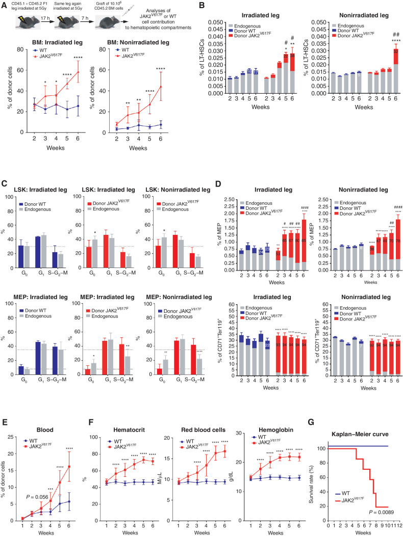
Figure 1.
Follow up of PV development in mice. A, Experimental design (top) and kinetics of transplanted WT or JAK2V617F BM cells contribution to total BM hematopoietic cells in the irradiated and nonirradiated legs (bottom). B, Kinetics of WT or JAK2V617F cells contribution to the long-term hematopoietic stem cell (LT-HSC). C, Cell-cycle analysis of WT (left, blue) and JAK2V617F (middle and right, red) Lin−cKit+Sca+ (LSK) cells (top) and megakaryocyte–erythroid progenitors (MEP, bottom) and their endogenous counterparts (grey) in the irradiated and nonirradiated legs 3 weeks after transplantation. D, Kinetics of WT or JAK2V617F cells contribution to MEP (top) and to CD71+Ter119+ mature erythroid (bottom) compartments in the irradiated and nonirradiated legs. E, Kinetics of peripheral blood cells after transplantation of JAK2V617F or WT BM cells in a single irradiated leg. F, Kinetics of blood parameters after transplantation of JAK2V617F or WT BM cells in a single irradiated leg. G, Survival curve of mice after transplantation of JAK2V617F or WT BM cells in a single irradiated leg. Data are mean ±SEM from three independent experiments (n = 8–12 mice). Significance was assessed using unpaired two-tailed t test and two-way ANOVA followed by post hoc analysis (*, P < 0.05; **, P < 0.01; ***, P < 0.001; ****, P < 0.0001; B and D, asterisks denote P-value comparing chimerism (% of GFP cells as indicated with the number (mean)) between Donor JAK2 vs Donor WT, and # denotes P-value comparing the % of cells studied from BM (irradiated and nonirradiated leg) of mice transplanted with WT or JAK2V617F cells).

