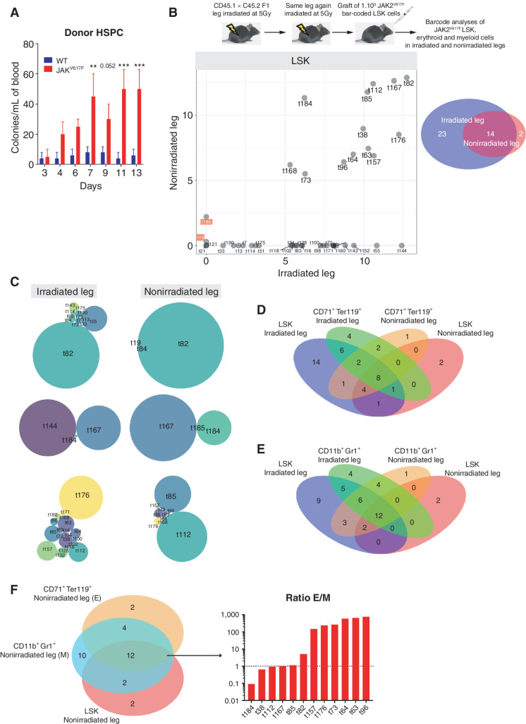
Figure 2.
JAK2V617F LSK from irradiated leg colonize the nonirradiated leg. A, Kinetics of donor-derived WT or JAK2V617F colony-forming unit per mL of blood from mice transplanted with WT or JAK2V617F BM cells in a single irradiated leg. Data are mean ±SEM. Significance was assessed using unpaired two-tailed t test and two-way ANOVA followed by post hoc analysis (**, P < 0.01; ***, P < 0.001). B, Experimental design (top) and frequency of JAK2V617F LSK bar-coded clones in the nonirradiated (y-axis) and irradiated (x-axis) legs (bottom left), the barcode frequency is log transformed and bar-coded clones discussed in the main text are highlighted. Venn diagram of bar-coded JAK2V617F LSK clones in the nonirradiated and irradiated legs (n = 3, bottom right). C, Representative distribution of bar-coded JAK2V617F LSK clones in the nonirradiated and irradiated legs for each mouse (n = 3). D, Venn diagram of bar-coded JAK2V617F LSK and CD71+Ter119+ erythroid clones in the nonirradiated and irradiated legs (n = 3). E, Venn diagram of bar-coded JAK2V617F LSK and CD11b+Gr1+ myeloid clones in the nonirradiated and irradiated legs (n = 3). F, Venn diagram of bar-coded JAK2V617F LSK, CD71+Ter119+ erythroid and CD11b+Gr1+ myeloid clones in the nonirradiated leg (left) and histograms showing the ratio of distribution of erythroid (E) and myeloid (M) bar-coded clones (right).

