Figure 1.

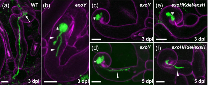
Infections formed by the succinoglycan‐deficient exoY mutant and the exoHKdel/exsH‐1345 mutant on Medicago truncatula Jemalong sunn‐2.
(a) WT Sinorhizobium meliloti 1021 expressing cCFP in a fully extended infection thread formed at 3 dpi on M. truncatula Jemalong sunn‐2. (b–d) CCRHs and short epidermal infection threads formed by the S. meliloti exoY mutant also expressing cCFP at 3 dpi (b, c) or 5 dpi (d). Note that in (c) and (d) the same site was imaged at two time‐points. (e, f) Infections formed by the exoHKdel/exsH‐1345 mutant at 3 dpi (e) or 5 dpi (f). Short, aborted infection threads (b, d, f) are labeled with arrowheads. The microcolony (infection chamber) is indicated in (a) (arrow). Excessively large root hair microcolonies (b–e) are labeled with asterisks. Images in (a–f) are z‐projections of confocal image stacks, combining the cCFP fluorescence of bacteria (green) and root hair cell wall autofluorescence (magenta). Results presented are representative of seven infection sites for S. meliloti WT 1021 (a), 24 sites for S. meliloti exoY (b–d) and 31 sites for S. meliloti exoHKdel/exsH‐1345 (e, f) recorded on three plants (six roots), five plants (11 roots) and seven plants (11 roots) respectively. Scale bars: 10 μm.
