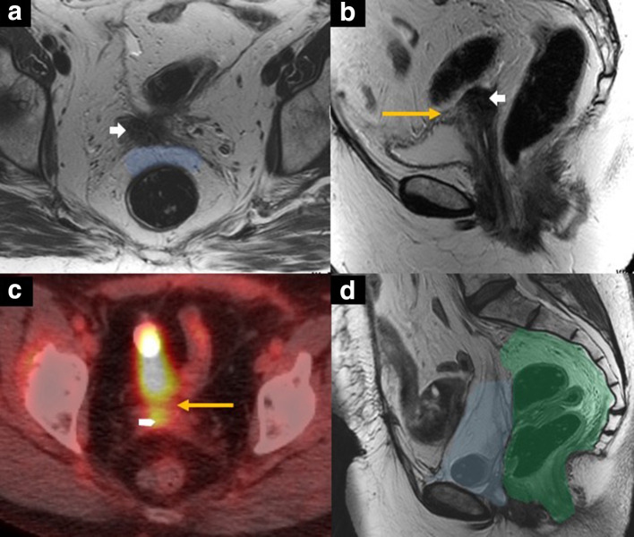
Figure 10.
60-year-old female with central pelvic recurrence of cervical cancer after radical hysterectomy and pelvic radiation therapy. Axial T2 weighted MR image (A) showing nodular mass (white arrow in A and B) at the vaginal cuff. There is a well-defined preserved plane of mesorectal fat between the mass and the rectum (blue-shaded area in A). Sagittal T2 weighted MR image (B) showing broad-based abutment and tethering of the posterior wall (yellow arrow in B) by the mass (white short arrow in B). On 18FDG PET/CT (C), the mass is hypermetabolic (white short arrow in C), and the posterior bladder focal abutment is again seen (yellow arrow in C). Sagittal T2 weighted MR image (D) after anterior pelvic exenteration with ileal conduit urinary diversion showing the postoperative situs with bowel loops and mesentery filling the former vesical space (blue-shaded area in D). The posterior compartment with the rectum and the anal sphincter complex (green shaded area in D) were spared.

