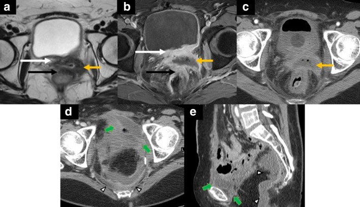
Figure 11.
52-year-old female with central pelvic recurrence of cervical cancer after radical hysterectomy and adjuvant chemoradiation therapy. Axial T2 weighted MR image (A) showing an intermediate signal irregular mass at the vaginal cuff (yellow arrow in A) with abutment and tethering of the posterior bladder wall (white arrow in A) and abutment of the anterior rectal wall without preserved fat planes (black arrow in A). Axial fat-suppressed post-contrast T1 weighted MR image (B) depicts the centrally necrotic mass (yellow arrow in B). Note the thickened posterior bladder wall (white arrow in B) and the anterior rectal wall (black arrow in B), suggesting invasion. Axial contrast-enhanced CT image (C) depicts the rim-enhancing, centrally necrotic mass with central foci of gas (yellow arrow in C). The patient underwent a total pelvic exenteration since the tumor involved the anterior and posterior compartments. Axial (D) and sagittal (E) contrast-enhanced CT images showing the postoperative situs complicated by rim-enhancing fluid-collections in the retropubic (short green arrows in D, E) and presacral space (white arrowheads in D, E) consistent with abscess collections.

