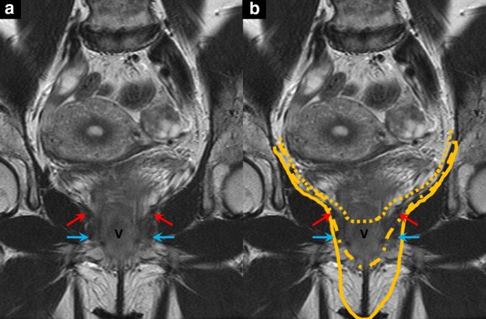
Figure 2.
Coronal T2 weighted MR image (A) with the arrows indicating the following anatomical landmarks: levator ani with red arrows, urogenital diaphragm with blue arrows. V = vagina. Image (B) shows the different resection planes of a supralevator and infralevator exenteration. The dotted line indicates a supralevator exenteration, the dash-dotted line an infralevator exenteration, and the solid line an infralevator exenteration with vulvectomy.

