Figure 6.
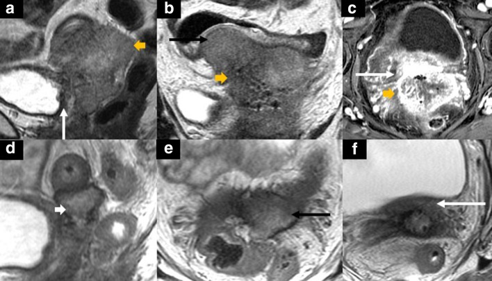
48-year-old female with recurrent cervical squamous cell carcinoma. Contrast-enhanced MRI of the pelvis at baseline (A: sagittal T2 weighted; B: coronal T2 weighted; C: axial fat-suppressed contrast-enhanced T1 weighted) and T2 weighted MR images after chemotherapy (sagittal [D] and axial [E-F]). There is a solid mass (yellow short arrow in A-C) centered in the pelvis involving the sigmoid colon (black arrows in B) and posterior bladder wall (white arrows in A and C). After chemotherapy, there is a marked reduction in tumor size (white short arrow in D) with concomitantly decreased involvement of the sigmoid wall (black arrow in E) and the posterior bladder wall (white arrow in F). She subsequently underwent a complete infralevator exenteration with vaginectomy with an end colostomy, ileal conduit, and right-sided VRAM flap reconstruction.

