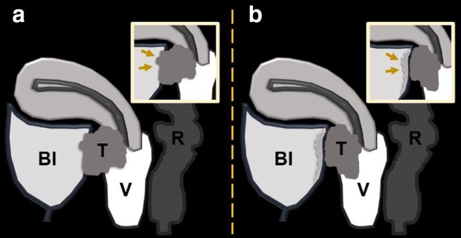
Figure 8.

Schematic illustration of a sagittal image with a cervical tumor (T) extending to the posterior bladder wall (Bl) with frank invasion in A (arrows in the magnified image in A) and abutment of the posterior bladder wall in B with reactive or inflammatory bullous edema of the posterior bladder wall (arrows in the magnified image in B). Please note that the signal of bullous edema is different from the signal of the primary tumor. R = rectum, V = vagina.
