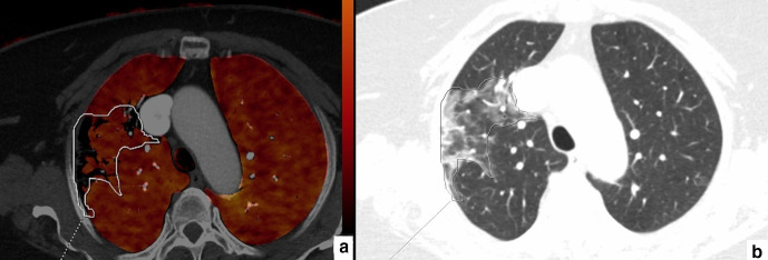
Figure 3.
64-year-old female. Axial perfusion blood volume (a) and computed tomography (b) images. In the right upper lobe, a large, heterogeneous area of GGOs and consolidations can be seen (b, marked area). Perfusion deficits are also present in the same area, but they take up less space than GGOs and consolidations (a, marked area).

