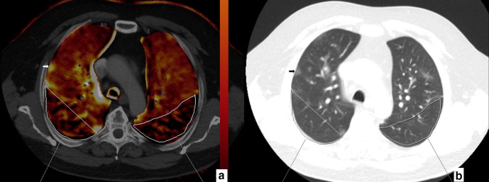
Figure 4.
67-year-old male. Axial perfusion blood volume (a) and computed tomography (b) images. Large, heterogeneous perfusion deficit areas are seen in the posterior zones of upper lobes (a, marked areas). No GGO or consolidation is present in the perfusion deficit areas (b, marked areas). Ground glass opacification in the right upper lobe peripheral zone (b, arrow) does not reveal any perfusion deficit (a, arrow).

