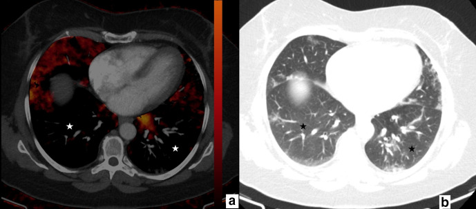
Figure 5.
77-year-old male. Axial perfusion blood volume (a) and computed tomography (b) images. Peripherally distributed ground glass opacifications and focal consolidations are present (b), large areas of perfusion deficits in the bilateral lower lobes are present on perfusion blood volume images (a, stars). Perfusion deficit areas occupy a larger area than GGOs and consolidations and mainly appear normal on computed tomography (b, stars).

