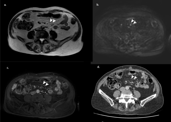
Figure 1.
MRI staging example of an 80-year-old female after three courses of neoadjuvant chemotherapy. T2 weighted (a) shows a suspicious dark thickening of the mesenteries (arrowheads), which showed diffusion restriction and contract enhancement on b1000 diffusion-weighted (b) and gadolinium-enhanced T1 weighted (c) imaging. At CRS, millimetric depositions were found on the mesenteric surface. A CT of 3 weeks earlier showed a similar pattern (arrowheads); however, with CT alone, no confident distinction can be made between ascites, fibrosis or PM. Therefore, the involvement of the mesenteries was not mentioned in the original CT report. CRS, cytoreductive surgery.

